B-h4-1BB/hHER2 mice
| Strain Name |
C57BL/6-Tnfrsf9tm1(TNFRSF9)Bcgen Erbb2tm1(ERBB2)Bcgen/Bcgen |
Common Name | B-h4-1BB/hHER2 mice |
| Background | C57BL/6 | Catalog number | 111869 |
|
Related Genes |
TNFRSF9 (tumor necrosis factor receptor superfamily, member 9); ERBB2, CD340, HER-2, HER-2/neu, HER2, MLN 19, NEU, NGL, TKR1, VSCN2 |
||
mRNA expression analysis

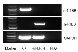
Strain specific analysis of 4-1BB gene expression in wild type (WT) mice and B-h4-1BB/hHER2 mice by RT-PCR. Mouse Tnfrsf9 mRNA was detectable only in spleen of WT mice (+/+). Human TNFRSF9 mRNA was detectable only in homozygous B-h4-1BB/hHER2 mice (H/H;H/H) but not in WT mice (+/+).

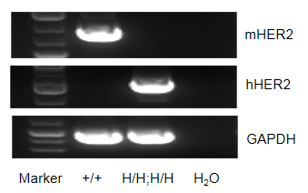
Strain specific analysis of HER2 gene expression in wild type (WT) mice and B-h4-1BB/hHER2 mice by RT-PCR. Mouse Erbb2 mRNA was detectable only in liver of WT mice (+/+). Human ERBB2 mRNA was detectable only in homozygous B-h4-1BB/hHER2 mice (H/H;H/H) but not in WT mice (+/+).

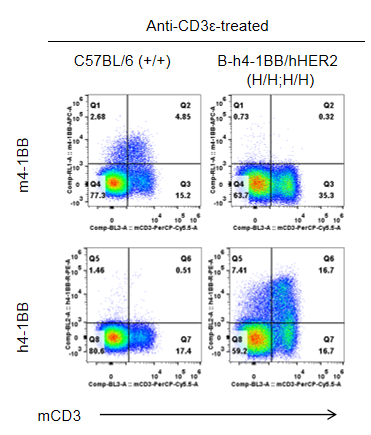
Strain specific 4-1BB expression analysis in homozygous B-h4-1BB/hHER2 mice by flow cytometry. Splenocytes were collected from wild type (WT) mice (+/+) and homozygous B-h4-1BB/hHER2 mice (H/H;H/H) stimulated with anti-CD3ε in vivo, and analyzed by flow cytometry with species-specific anti-4-1BB antibody. Mouse 4-1BB was detectable in WT mice (+/+). Human 4-1BB was exclusively detectable in homozygous B-h4-1BB/hHER2 mice (H/H;H/H) but not in WT mice (+/+).

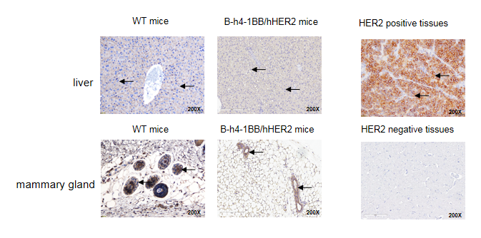
Immunohistochemical (IHC) analysis of HER2 expression in homozygous B-h4-1BB/hHER2 mice. The liver and mammary gland were collected from WT mice and homozygous B-h4-1BB/hHER2 mice and analyzed by IHC with anti-HER2 antibody. HER2 was detectable in WT mice and homozygous B-h4-1BB/hHER2 mice due to the cross-reactivity of the antibody. The arrow indicates tissue cells with positive HER2 staining (brown).


