• 311445
The mouse Cd38 gene was replaced by chimera CDS composed of human CD38 extracellular coding sequence plus mouse Cd38 cytoplasmic sequence and the full coding sequence of luciferase in B-hCD38-Luc E.G7-OVA cells. Human CD38 was highly expressed on the surface of B-hCD38-Luc E.G7-OVA cells.
Gene targeting strategy for B-hCD38-Luc E.G7-OVA cells. The mouse Cd38 gene was replaced by chimera CDS composed of human CD38 extracellular coding sequence plus mouse Cd38 cytoplasmic sequence and the full coding sequence of luciferase in B-hCD38-Luc E.G7-OVA cells. The replacement sequences disrupts the endogenous murine Cd38 gene, resulting in a non-functional transcript.
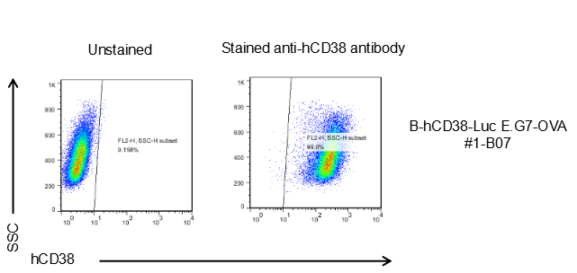
CD38 expression analysis in B-hCD38-Luc E.G7-OVA cells by flow cytometry. Single cell suspensions from B-hCD38-Luc E.G7-OVA cultures were stained with species-specific anti-hCD38 antibody. Human CD38 was detected on the surface of B-hCD38-Luc E.G7-OVA cells. The 1-B07 clones of B-hCD38-Luc E.G7-OVA cells can be used in vivo experiments.
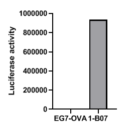
Luciferase reporter activity assay of B-hCD38-Luc E.G7-OVA cells. Single cell suspensions from B-hCD38-Luc E.G7-OVA cultures were collected. The luciferase activities were measured by Bright-GloTM luciferase Assay System (Promega, Cat E2610), and B-hCD38-Luc E.G7-OVA cells have strong luminescence signal. The 1-B07 clones of B-hCD38-Luc E.G7-OVA cells can be used in vivo experiments.
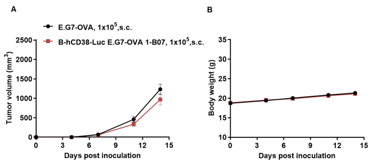
Subcutaneous homograft tumor growth of B-hCD38-Luc E.G7-OVA cells. B-hCD38-Luc E.G7-OVA cells (1x10⁵) and wild-type E.G7-OVA cells (1x10⁵) were subcutaneously implanted into C57BL/6 mice (female, 7-week-old, n=5). Tumor volume and body weight were measured twice a week. (A) Average tumor volume ± SEM. (B) Body weight (Mean± SEM). Volume was expressed in mm3 using the formula: V=0.5 X long diameter X short diameter2. As shown in panel A, B-hCD38-Luc E.G7-OVA cells were able to establish tumors in vivo and can be used for efficacy studies.
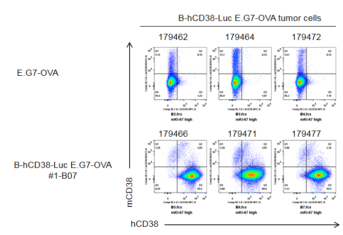
B-hCD38-Luc E.G7-OVA cells were subcutaneously transplanted into C57BL/6 mice (n=5). At the end of the experiment, tumor cells were harvested and assessed for human CD38 expression by flow cytometry. As shown, human CD38 was highly expressed on the surface of tumor cells. Therefore, B-hCD38-Luc E.G7-OVA cells can be used for in vivo efficacy studies of novel CD38 therapeutics.
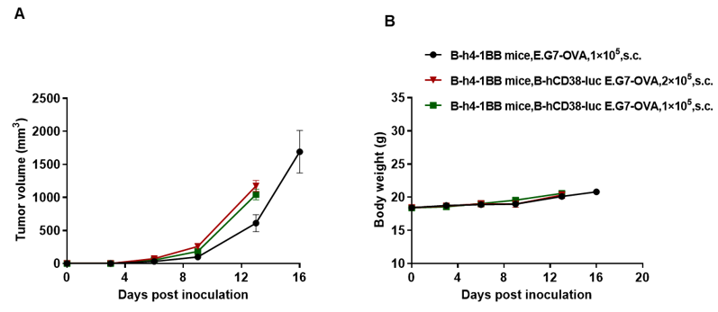
Subcutaneous homograft tumor growth of B-hCD38-luc E.G7-OVA cells. B-hCD38-luc E.G7-OVA cells and wild-type E.G7-OVA cells were subcutaneously implanted into B-h4-1BB mice (female, 7-week-old, n=6). Tumor volume and body weight were measured twice a week. (A) Average tumor volume. (B) Body weight. Volume was expressed in mm3 using the formula: V=0.5 X long diameter X short diameter2. As shown in panel A, B-hCD38-luc E.G7-OVA cells were able to establish tumors in vivo and can be used for efficacy studies. Values are expressed as mean ± SEM.
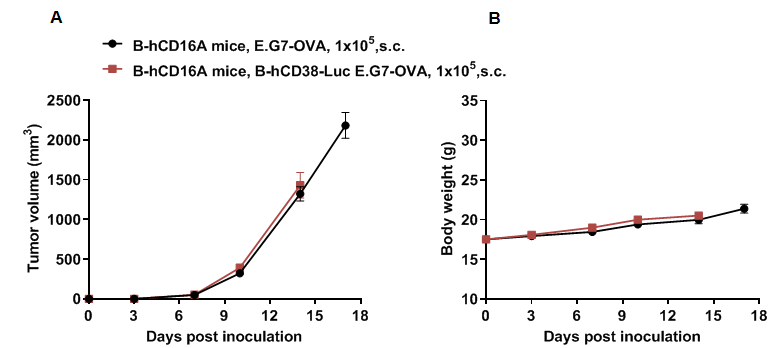
Subcutaneous homograft tumor growth of B-hCD38-Luc E.G7-OVA cells. B-hCD38-Luc E.G7-OVA cells (1x10⁵) and wild-type E.G7-OVA cells (1x10⁵) were subcutaneously implanted into B-hCD16A mice (female, 6-week-old, n=8). Tumor volume and body weight were measured twice a week. (A) Average tumor volume ± SEM. (B) Body weight (Mean± SEM). Volume was expressed in mm3 using the formula: V=0.5 X long diameter X short diameter2. As shown in panel A, B-hCD38-Luc E.G7-OVA cells were able to establish tumors in vivo and can be used for efficacy studies.
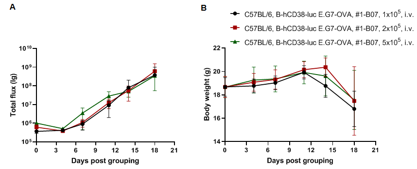
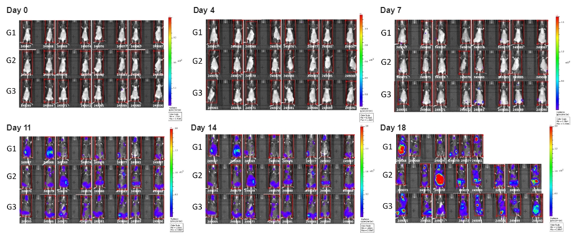
Tumor growth and in vivo imaging of B-hCD38-luc E.G7-OVA. B-hCD38-Luc E.G7-OVA cells were injected by tail vein into C57BL/6 mice. Signal intensity and body weight were measured twice a week. (A) Imaging was performed on days 0, 4, 7, 11 and 18, (B) Mice body weight (Mean ± SEM). B-hCD38-Luc E.G7-OVA cells can be used for in vivo efficacy evaluation.
In vivo luciferase images of B-hCD38-Luc E.G7-OVA cells